
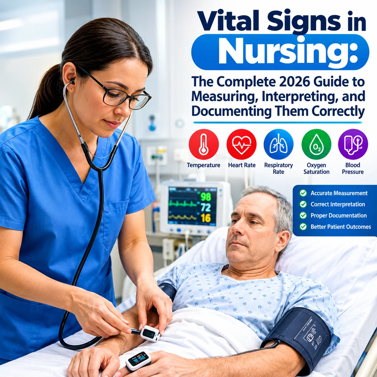
Vital Signs in Nursing: The Complete 2026 Guide to Measuring, Interpreting, and Documenting vital signs Correctly

Abdullahi Suleiman
September 13, 2026
The body can tell a story long before symptoms begin to speak for themselves. Vital signs are some of the earliest clues nurses use to understand what is happening within a patient’s body.
Vital signs in nursing are essential clinical measurements used to assess a patient’s health status. They serve as an objective foundation for clinical judgment and patient care planning. They provide information about a patient’s physiological status and can reveal early changes in the body. Errors in measurement or documentation may lead to poor clinical decisions and inaccurate assessment, affecting the entire nursing process.
This article will cover the major vital signs in nursing, how to measure vital signs correctly, how to document them, their normal ranges, common clinical mistakes, factors that can affect vital signs and more.
Table of Contents
- An Overview of What Are the 5 (or 6) Vital Signs in Nursing?
- What Are Vital Signs in Nursing?
- Importance of Vital Signs In Nursing
- How to Measure Vital Signs In Nursing
- Common vital signs route
- How to Record Vital Signs In Nursing
- Factors That Can Affect Vital Signs
- Common Mistakes While Measuring Vital Signs.
- When Should a Nurse Report Abnormal
- Tips for Accurate Vital Sign Measurement and Documentation
- FAQs About Vital Signs in Nursing
- Conclusion
An Overview of What Are the 5 (or 6) Vital Signs in Nursing?
- Temperature — normal adult range: 36.5–37.5°C (97.8–99.5°F)
- Pulse (Heart Rate) — normal adult range: 60–100 beats per minute
- Respiratory Rate — normal adult range: 12–20 breaths per minute
- Blood Pressure — normal adult range: below 120/80 mmHg
- Oxygen Saturation (SpO₂) — normal range: 95–100% on room air
- Pain — increasingly treated as the “6th vital sign,” rated on a 0–10 scale
Vital signs are checked on admission and repeated based on the patient’s condition, providing a baseline, an early warning system, and a way to track how a patient responds to treatment.
What Are Vital Signs in Nursing?
Vital signs are objective measurements used to assess a patient’s physiological function. They provide a baseline patient assessment and establish a foundation for the nursing process.
The major vital signs in nursing include temperature, pulse, respiratory rate, blood pressure and oxygen saturation.
Temperature
Body temperature shows the balance between heat production and heat loss in the body. Temperature is one of the core vital signs that can indicate changes in the body’s thermoregulatory function.
Body temperature can be influenced by environment, age, activity and other factors.
Pulse rate
Pulse rate is the number of times the heart beats per minute. It provides information about the heart rate, cardiac rhythm, circulation and peripheral tissue perfusion.
Nurses assess for rate, rhythm and volume
Rate: This is the total number of beats per minute
Rhythm: This refers to the pattern and regularity of the intervals between heartbeats. A regular rhythm has equal intervals between beats while an irregular rhythm has varying intervals and may indicate arrhythmia
Volume: This is the amplitude of the pulse felt in the artery. It can provide information about the strength of blood flow through the arterial system.
Abnormal changes in pulse rate should be assessed appropriately as they may be associated with physical exertion, fever, pain, medications, cardiac or circulatory problems.
Respiratory Rate
Respiratory rate is the number of breaths a person takes per minute. It is measured by counting the number of complete respiratory cycles with one inhalation and exhalation counted as one breath. Respiratory rate can be an early indicator of clinical deterioration because changes in breathing may occur when the body is trying to maintain adequate oxygenation and remove excess carbon dioxide.
Nurses don’t just assess the rate alone; they assess for depth, rhythm and effort.
Rhythm: Nurses assess whether the patient has a steady and consistent breathing pattern.
Depth: Nurses assess the amount of air moving in and out of the lungs as indicated by the depth of the chest movement. It ranges from shallow to deep.
Effort: This refers to the work required to breathe. Normal breathing is generally automatic, silent and unlabored. Nurses assess for signs of increased work of breathing such as labored breathing, use of accessory muscles, wheezing, and nasal flaring.
For example,
A patient experiencing an asthma attack may have prolonged or difficult expiration and wheezing. The patient may also take deeper or faster breaths as the body attempts to meet its oxygen demands.
READ ASLO: Nurses on Social Media: Digital Literacy, Professionalism
Blood Pressure (BP)
This is the force of blood pushing against the walls of the arteries as the heart pumps blood through the body. It is measured in millimeters of mercury (mmHg) and recorded as systolic and diastolic pressure. The systolic pressure is the top number while the diastolic is the bottom number i.e 120/80 mmHg
Systolic pressure measures the pressure in the artery when the heart contracts and pumps blood out.
Diastolic pressure measures the pressure in the artery when the heart relaxes between beats.
Monitoring blood pressure is essential because it helps assess the cardiovascular function, detects hypertension (high BP) or hypotension (low BP), monitor changes in a patient’s condition and guide treatment decisions.
Oxygen Saturation
Oxygen saturation is the percentage of hemoglobin in the blood that is carrying oxygen. It is usually measured using a pulse oximeter, which is commonly placed on the finger, toe or earlobe.
Oxygen saturation helps nurses assess how effectively the lungs are oxygenating the blood and whether the body is receiving adequate oxygen.
Interpretation of the patient’s oxygen saturation differs with patient. While 90% may be concerning in some patients, a COPD patient might have a different prescribed target.
Therefore, oxygen saturation should be interpreted alongside the patient’s baseline, clinical condition and prescribed oxygen target where applicable.
Importance of Vital Signs In Nursing
Vital Signs in nursing are one of the core foundations of nursing practice because it provides information on a patient’s physiological condition.
Vital signs are taken on admission and repeated based on the patient’s condition to monitor changes, guide care plan and identify problems.
Vital signs helps nurses to
- Establish a baseline for the patient
Vital signs serves a benchmark for establishing a patient’s baseline and for comparing future measurements. A baseline helps nurses to determine what is normal for a patient based on the initial assessment and clinical condition.
- Detecting deterioration
Vital signs serve as warning signs of abnormalities in a patient’s health status. Nurses must recognize this warning signs and respond to it appropriately.
- Monitoring a patient’s treatment
Vital signs help recognize if a patient is responding effectively to treatment.
- Communicating patient’s health status to the health care team.
Well-documented vital signs help the healthcare team to understand the condition of a patient and identify changes over time. They also assists nurses in triaging patients and prioritizing care based on urgency.
How to Measure Vital Signs In Nursing
Temperature
Measuring temperature accurately requires the right equipment, considering the route and proper documentation. The normal range for calculating temperature is 36.5-37.5°C and it varies by hospital, route or the patient’s condition. It is also measured in Fahrenheit.
Equipment for measuring temperature includes thermometer, cotton wool swab, gloves when necessary.
Patient preparation
Confirm the patient’s identity
Explain the procedure and seek consent.
Inquire about any recent activity of the patient that may affect the reading.
Perform hand hygiene and wear gloves where necessary.
Note that the patient shouldn’t partake in any activity like exercising, consuming things that are either hot or cold that may affect the reading about 25-30mins before.
Common vital signs route
Oral route
The thermometer is placed in a posterior sublingual position under the tongue. The patient closes their mouth around the thermometer.
Thermometer should not be used for an unconscious patient, an uncooperative patient and children because of risk of biting. Clean the thermometer according to the institution policy. This route could be limited by food substances in the mouth, mouth breathing, smoking or inability to cooperate. Normal ranges may vary depending on the measurement route, the patient’s condition, and the reference range used by the healthcare facility.
Axillary route
The thermometer is placed in the middle of the armpit and held firmly by the arm. This requires proper positioning of the thermometer in the armpit and may be affected by room temperature.
Tympanic route
The thermometer is placed gently in the ear canal and aimed at the ear drum. Tympanic thermometer measures infrared heat from the tympanic membrane. This could be limited by incorrect positioning, earwax, infection and more. Avoid pushing it into the ear with force and it is advisable to be done under supervision.
Rectal route
A lubricated thermometer is placed inside the rectum and the depth is determined by the device instruction or clinical policy. It is usually accurate but should be avoided when rectal surgery has taken place, cases whereby the patient has diarrhea, infection or if it is against the hospital policy.
Other thermometers include;
Infrared temperature: This measures infrared heat emitted from the body without direct contact and could be affected by technique or environmental factors.
Analog thermometer: This is a glass thermometer that uses liquid to indicate temperature on a scale. It is read at eye level.
How to use Analog Thermometer
Clean the thermometer with cotton wool swab from bottom to top.
Shake it down until the liquid is below the temperature mark
Explain the procedure to the patient and put it in the appropriate route.
Leave it for a specified time commonly a few minutes. Some may sound after reading.
Remove it and read it at eye level.
Clean the thermometer
Document showing the time, route, date.
Factors affecting temperature includes age, time, exercise, emotional stress, dehydration, recent bathing, pregnancy, menstruation, specific medications and many more.
Pulse
Pulse assessment measures the heart rate and provides information about circulation. The radial artery is the common site for pulse assessment. It is measured by placing the index finger and middle finger over the radial artery and applying gentle pressure until the pulse is felt. Don’t use the thumb because it has its own palpable pulse which can interfere with the assessment. Pulse rate is recorded in beats per minute (bpm). For an adult at rest, the normal range is 60 – 100 bpm. Although the pulse may be counted for 30 seconds and multiplied by two when the rhythm is regular, counting for a full 60 seconds is recommended when the rhythm is irregular.
While counting, it is necessary to assess whether the pulse is faint or strong and whether it is regular or irregular.
Other pulse assessment sites include:
Brachial artery — located on the inner aspect of the upper arm, commonly used for blood pressure measurement and in infants.
Carotid artery — located on the side of the neck.
Popliteal artery — located behind the knee.
Femoral artery — located in the groin.
And others.
Respiratory rate
This is measured by observing the patient’s breathing pattern and counting the number of breaths taken in one minute with inhalation and exhalation counted as one respiration. Observe the movement from the chest or abdomen.
When possible, count the respiration without drawing the patient’s attention to the fact that you are counting their breathing, as they may become conscious of their breathing and unconsciously alter their breathing pattern.
While counting the respiratory rate, assess the regularity of the breathing, the depth of the breathing and the amount of effort required to breathe.
It is also necessary to observe for any form of respiratory distress like rapid or slow breathing, chest retraction, wheezing, use of accessory muscles, difficulty speaking or breathing properly, cyanosis and other abnormalities.
If respiratory distress is suspected, seek assistance from a senior nurse/ any appropriate healthcare practitioner.
Blood pressure
Accurate measurement requires proper positioning, correct cuff placement and an appropriate rest period. Small errors in technique can affect the accuracy of the blood pressure reading.
Before measuring the blood pressure, the patient should avoid exercise, caffeine and smoking for about 30 minutes when possible. The patient should also empty their bladder and rest for at least 5 mins before taking the measurement.
The patient should be seated comfortably in an upright position, with their back supported, feet flat on the floor, and legs uncrossed. The arm should be bare, supported, and positioned at heart level. For patients who cannot sit or must remain in bed, blood pressure may be measured in a lying or other appropriate position, with the arm supported at heart level as much as possible. The patient should not hold the arm up, clench the fist, or leave the arm unsupported. The patient should also avoid talking during the measurement.
Using the Automated Blood Pressure Monitor
The automated blood pressure monitor is easier to use and does not require a stethoscope.
Place the cuff around the patient’s upper arm with the lower edge above the antecubital fossa and the artery marker aligned with the brachial artery. Following the manufacturer’s instruction, press the start button and allow the monitor to complete the measurement.
Ensure the cuff is worn properly and is of appropriate size for the patient’s arm circumference. A cuff that is too small may produce a falsely high reading, while one that is too large may produce a falsely low reading.
Using a manual sphygmomanometer
Place the stethoscope over the brachial artery without pressing excessively. Inflate the cuff until the pulse is no longer palpable and then slightly further. Deflate the cuff slowly and steadily while listening for Korotkoff sounds. The pressure at which the first clear korotkoff sounds are heard represents the systolic pressure. The pressure when the sounds disappear represents the diastolic pressure.
Range for Blood Pressure
| Blood pressure category | Systolic BP (mmHg) | Diastolic BP (mmHg) |
| Normal | <120 | <80 |
| Elevated | 120-129 | <80 |
| Hypertension stage 1 | 130-139 | 80-89 |
| Hypertension stage 2 | >140 | >90 |
| Hypertensive crisis | >180 | >120 |
N.B Normal ranges may vary depending on the patient’s condition, baseline and the reference range used by the healthcare facility.
Oxygen Saturation
Oxygen saturation is measured using a pulse oximeter. The device is commonly placed on the finger although other sites like earlobe or toe could be suitable. The site should be warm, clean and have adequate circulation.
When reading the oxygen saturation of the patient, position the light emitter and sensor correctly to avoid inaccurate reading. Allow the patient hands to relax and wait until the displayed oxygen saturation stabilize and stop changing values.
The normal range for oxygen saturation is 95-100% but may differ with the patient’s condition. Hence, oxygen saturation should be interpreted alongside the patient’s condition.
Factors that affect inaccurate reading include poor peripheral circulation, low blood pressure, incorrect placement or size of the device, cold extremities and others.
How to Record Vital Signs In Nursing
Recording vital signs is quite different from measuring vital signs. Recording vital signs simply means documenting the measurement obtained. It is useful for communicating changes, evaluating treatment and identifying trends.
Documentation may differ according to the hospital’s policy. Some hospitals may use one device or route for measuring vital signs. Ranges may also differ depending on the institution. Therefore, documentation should follow institutional policies and guidelines.
Vital signs must be:
Accurate: Documentation should be precise and free from errors. Record the values as measured without altering or guessing the result.
Timely: Record the vital signs in the medical record as soon as the measurement is taken to show the patient’s current physiological status and to identify trends. While recording, it is necessary to note down the time the measurement was taken.
Complete: A vital sign documentation must be complete. It should include all the required measurements, abnormal findings, interventions and other information required by the institution.
Legible and Clear: Use approved clinical abbreviations. For electronic health records [EHR], ensure information is entered properly and in the appropriate field.
Consistent with the facility’s documentation system: Follow the institution’s guidelines for time format, authentication, observation chart, flow sheet and procedures for documenting abnormal readings.
WHAT TO DOCUMENT
Date
Time
Vital signs values
Route/site where applicable
Patient’s position
Relevant observations
Oxygen therapy where applicable
Nursing interventions when applicable.
An Example
08 Aug 2026, 9:52 a.m.: T 37.6°C (axillary), P 72 bpm, RR 20 cpm, BP 121/76 mmHg, SpO₂ 96% on room air. Patient positioned in semi-Fowler’s position. No signs of respiratory distress noted.
Factors That Can Affect Vital Signs
Exercise
Age
Body position
Anxiety
Pain
Recent meal
Environment
Dehydration
Medications
Fever or Infection
Caffeine and other stimulants.
Common Mistakes While Measuring Vital Signs.
- Using the wrong blood pressure cuff size:
Using a cuff that is too small may produce a falsely high reading while one that is too large may produce a falsely low reading. It is important to use a cuff that is appropriate for the patient’s arm circumference.
- Measuring BP without adequate rest:
Inadequate rest may elevate the reading and lead to inaccurate assessment. When clinically appropriate, the patient should rest quietly for about 5 minutes before measurement.
- Counting respiration inaccurately:
The patient may alter their breathing when they realize that it is being observed. When possible, observe and count the patient’s respirations without drawing attention to the fact that their breathing is being counted.
- Ignoring pulse rhythm or quality:
This may lead to missed signs of irregular rhythm or weak pulse and can delay the recognition of conditions that require further assessment.
- Failing to report significant abnormalities:
Documentation alone doesn’t guarantee prompt intervention. When significant abnormalities are identified, the nurse should assess and reassess the patient, document the findings and notify the appropriate healthcare professional or senior nurse according to institutional policy.
When Should a Nurse Report Abnormal
Vital Signs?
Identifying an abnormal vital sign is a nursing responsibility. Knowing when to escalate it requires clinical judgement. Vital-sign measurements and monitoring may differ between patients depending on their age, condition, baseline and clinical situation. When an abnormal reading is identified, compare it with the previous measurements, the patient’s baseline, history and other factors that may have actually contributed to it. However, significant abnormalities associated with signs of deterioration should be escalated promptly.
What is normal for one patient may be abnormal for another.
Another risk factor that should be considered is when there is a continuous decline in the series of measurement.
Institutions are provided with facility escalation protocols which provide clear instructions on when to notify a doctor or a rapid response team(RRT)
Read Also: Overview of Nursing Documentations Practice for Blood Transfusion Reaction
Tips for Accurate Vital Sign Measurement and Documentation
To obtain accurate measurement and documentation of the patient, it is necessary for the nurse to:
Confirm the identity of the patient
Explain the procedure to the patient.
Perform hand hygiene before and after procedure.
Use the correct and appropriate equipment.
Ensure the equipment is clean and functioning properly.
Position the patient properly when possible.
Take measurements carefully and accurately.
Reassess the patient when an abnormal measurement is obtained.
Compare the current reading with previous measurement and patient’s baseline.
Record findings promptly and accurately.
Report concerning changes according to the institution policy.
FAQs About Vital Signs in Nursing
What are the normal vital signs for adults?
Normal vital signs can vary depending on age, clinical condition and institution protocols. For a resting adult, the commonly use reference range are:
Temperature: 36.5-37.5°C
Pulse rate: 60-100 beat/minute (bpm)
Respiratory rate: 12-20 breaths/ minute
Blood pressure: <120/80 mmHg
Oxygen saturation: 95-100%
Note that these values should not be interpreted in isolation. The patient’s baseline, previous measurements, clinical condition and assessment findings should be considered.
How often should vital signs be checked?
There is no fixed frequency for checking vital signs. They should be checked according to the patient’s stability, clinical setting and specific medical orders. Unstable patients may require more monitoring.
Why do we avoid placing the cuff over loose clothing?
Clothing and poor cuff contact can interfere with the pressure transmitted to the arm. The cuff may not sit properly over around the arm and align correctly with the brachial artery which can lead to inaccurate measurement.
How does eating affect the measurement of blood pressure?
Eating a meal shortly before measuring the blood pressure may affect the reading because digestion may alter the blood flow. When possible, the blood pressure should be measured in the patient’s rest state and according to recommended measurement guidelines.
How often should vital signs be checked? There’s no fixed universal frequency — vital signs should be checked according to the patient’s stability, the clinical setting, and specific medical orders. Unstable or critically ill patients require more frequent monitoring than stable ones.
Why is blood pressure cuff size important? A cuff that’s too small produces a falsely high reading, while one that’s too large produces a falsely low reading. The cuff bladder should encircle roughly 80% of the patient’s arm for an accurate result.
Why do nurses avoid measuring blood pressure over clothing? Clothing and poor cuff contact interfere with how pressure transmits to the arm, preventing proper alignment with the brachial artery and leading to inaccurate readings.
Does eating affect blood pressure measurement? Yes. Eating shortly before a blood pressure check can alter blood flow through digestion, potentially skewing the reading — measurements should ideally be taken with the patient at rest, following standard guidelines.
Which vital sign changes first when a patient is deteriorating? Respiratory rate is widely recognized as one of the earliest and most sensitive indicators of clinical deterioration, often changing before other vital signs become abnormal.
What should a nurse do if a vital sign reading looks wrong? Never dismiss an unusual reading — re-check it, ideally with a second method or site, and assess the patient for any signs consistent with that value. When in doubt, ask a colleague to verify the measurement before acting on it.
Conclusion
Vital signs are more than mere numbers recorded on a chart. They provide information about a patient’s physiological status and help nurses recognize changes in the patient’s health condition. Mastering vital signs in nursing means understanding how to measure and record vital signs accurately helps nurses and other health practitioners establish a reliable baseline, identify deterioration and communicate changes promptly. This also supports clinical decision-making and patient care.
Vital signs are a critical part of professional nursing practice and directly impacts patient outcomes when accurately measured, interpreted, documented and acted upon.
Writing by Ayilara Oluwadunmininu
Health content writer